GY-5200 stereo electrocardiograph
Classification:
Product Introduction
Product Details
The stereo electrocardiograph supports Wilson and Frank lead systems and can collect regular 12-lead, 15-lead, 18-lead and orthogonal lead electrocardiograms. The stereo electrocardiograph also supports the synchronous acquisition of these two lead systems. One acquisition can obtain stereo electrocardiogram (vector), twelve-lead electrocardiogram, twelve-lead electrocardiogram, right chest lead or posterior wall lead electrocardiogram, orthogonal electrocardiogram, time electrocardiogram vector map, continuous electrocardiogram vector map, variable electrocardiogram vector map and other graphs.
The "secondary projection theory" in electrocardiography holds that the electrocardiogram is formed by the secondary projection of the three-dimensional ECG vector ring. However, it is easy to lose some important ECG information in the projection process, such as sudden steering, overlap, erosion and uplifting of the QRS vector ring, which is extremely important for the diagnosis of coronary heart disease, especially myocardial infarction. Stereo electrocardiogram makes up for these deficiencies in the diagnosis of regular electrocardiogram, eliminating the "blind area" of regular electrocardiogram and planar electrocardiogram and the overlapping part due to the angle of view. Therefore, when the ECG is suspicious or cannot be clearly diagnosed, it is necessary to make a three-dimensional ECG comparative analysis.
A large number of clinical studies have shown that stereotactic electrocardiography also has obvious advantages over regular electrocardiography in myocardial infarction complicated with bundle branch block, bundle branch block and multi-bundle branch block, ventricular hypertrophy, atrial arrhythmia with indoor differential conduction, diagnosis and differential diagnosis of ventricular arrhythmia, localization of the origin of ventricular arrhythmia (diagnosis and differential diagnosis of endocardial and epicardial ventricular premature beats), and diagnosis and localization of preexcitation syndrome.
The three-dimensional electrocardiograph produced by our company has designed a series of original functions according to the requirements of the majority of ECG doctors:
1. Up to 26 channels of ECG signal can be displayed and printed synchronously;
2. Unique heart beat number function, can choose heart beat analysis;
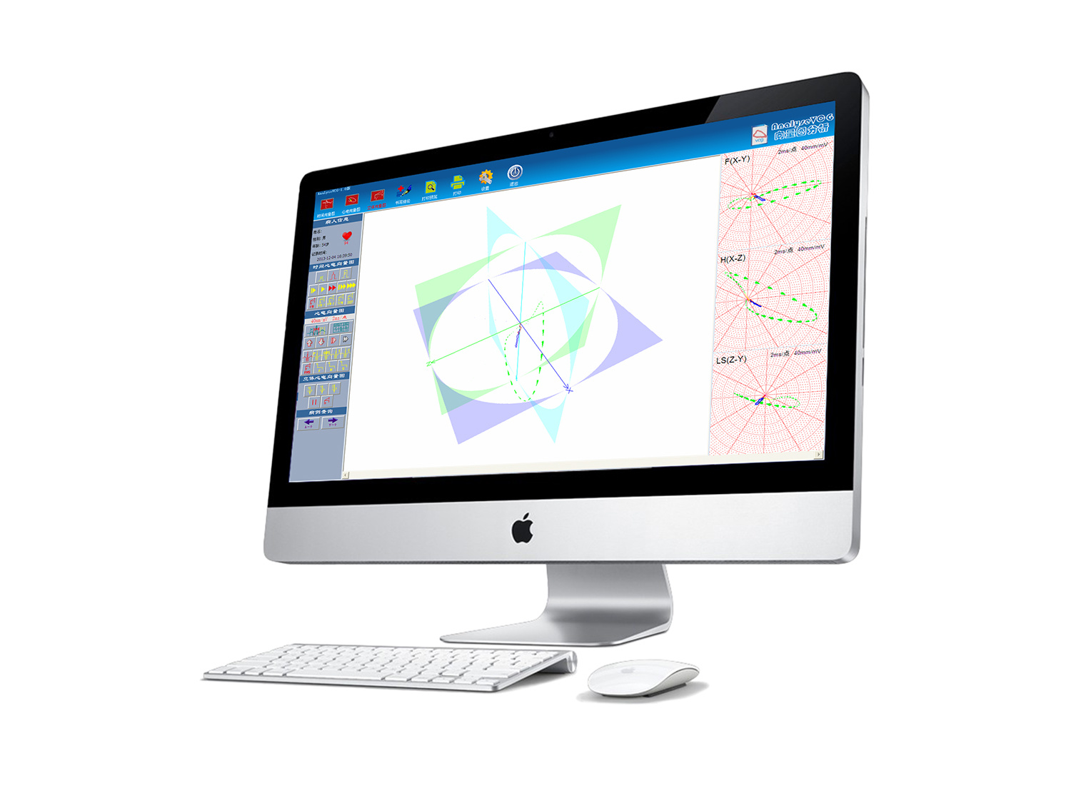
3. Overlapping printing function: ECG vector rings of multiple heart beats can be overlapped and displayed and printed, and the difference between normal and ectopic ECG vector rings (color display) can be clearly compared at a glance;
4. Color printing function: it supports different color display of P ring, QRS ring, T ring and QRS ring starting, middle and terminal parts respectively. The above functions can be customized;
5. Any angle can be 360 ° rotating color stereo vector ring body, support automatic rotation, mouse drag rotation;
6. Planar vectorcardiogram and stereo vectorcardiogram can be reconstructed after ordinary 12, 15 and 18 lead ECG acquisition;
7. Abundant parameter indicators to provide clinicians with more comprehensive diagnostic data;
8. Rich report templates for clinicians to choose freely;
The diagnostic criteria from the three-dimensional vectorcardiogram is accurate and unique, to achieve all-round, full-angle observation of the three-dimensional electrocardiogram, so that the diagnosis is more objective, comprehensive, detailed, intuitive, accurate and practical.
The three-dimensional vectorcardiogram analysis system realizes the omni-directional and full-angle observation of the electrocardiogram characteristics of atrial and ventricular depolarization and repolarization at all levels, which is helpful for the diagnosis of common cardiovascular diseases and the evaluation of the prognosis and risk of arrhythmia in patients.
The stereo electrocardiograph supports Wilson and Frank lead systems, and can replace the regular electrocardiograph for standard 12-lead, 15-lead, 18-lead and orthogonal lead electrocardiogram examinations in clinical applications. The stereo electrocardiograph also supports the synchronous acquisition of these two lead systems. One acquisition can obtain stereo electrocardiogram (vector), 12-lead electrocardiogram, 12-lead electrocardiogram right chest lead (V3R, V4R and V5R) or posterior wall lead (V7, V8 and V9) electrocardiogram, orthogonal electrocardiogram, time electrocardiogram, continuous electrocardiogram vector map, variable electrocardiogram vector map and other graphs. Three-dimensional electrocardiogram contains all the functions of ordinary electrocardiogram. Three-dimensional electrocardiogram can complete the work that ordinary electrocardiogram cannot complete, such as the overlapping function of multiple heart beats of the ECG vector ring described above, the running speed, direction and orientation of each part of the QRS ring, etc. These functions of the three-dimensional ECG instrument make it far beyond the regular ECG, and provide clinicians with a broader diagnosis and analysis of ideas.
As a multi-domain three-dimensional ECG (vector) map analysis and diagnosis integrated system, the three-dimensional ECG instrument contains a number of indicators and functions in the field of ECG diagnosis, which enables clinicians to evaluate the state of cardiac electrical activation and myocardial lesions in an all-round way, improves the sensitivity, specificity, accuracy and practicality of ECG diagnosis, and is more convenient for the classification, identification, diagnosis and treatment of clinical diseases.
Key words:
GY-5200 stereo electrocardiograph


